Alcon-TrueVision Partnership Delivers on Heads-Up Surgery
At OIS@ASRS 2016, Forrest Fleming, then CEO of TrueVision Systems, announced a new partnership with Alcon. He also introduced the result of that pairing: the Ngenuity 3D Visualization System for digitally assisted vitreoretinal surgery, featuring TrueVision’s heads-up, three-dimensional imaging technology delivered on Alcon’s platform. Fleming said it would be a platform that would “grow, and enhance, and change.”
Two years later, growth realized
That growth manifested at the 2018 meeting of the American Society of Retina Specialists (ASRS) in Vancouver when Alcon introduced its Ngenuity 3D Visualization System with Datafusion. The addition of the Datafusion software enables integration of Ngenuity with Alcon’s Constellation Vision System.
Alcon says this integration allows surgeons to track key data parameters in real time, such as intraocular pressure, flow rates, infusion pressure, and laser power, on one screen. Additional functionality simplifies the surgical experience allowing for customization, including the introduction of four preset imaging modes, two foot switch control options, advanced two- or three-dimensional video capture, and enhancement of the graphical user interface and procedure flow to provide an improved white balance process.
In a press release, Sergio Duplan, region president, North America, Alcon, stated, “We are combining two of our game-changing systems to streamline the customer experience, allowing surgeons to track real-time data feedback on one screen during procedures.”
Current TrueVision CEO Burton Tripathi, PhD, says that through the distribution agreement signed in 2016, Alcon became TrueVision’s exclusive marketing partner for its 3D technology in ophthalmology, at which time they rebranded the technology as Ngenuity. “We’re very happy with them,” Dr. Tripathi says. “They are doing a great job building awareness and growing the market.” As for TrueVision, he adds, the company continues to grow, and revenues are up year after year.
The Ngenuity System
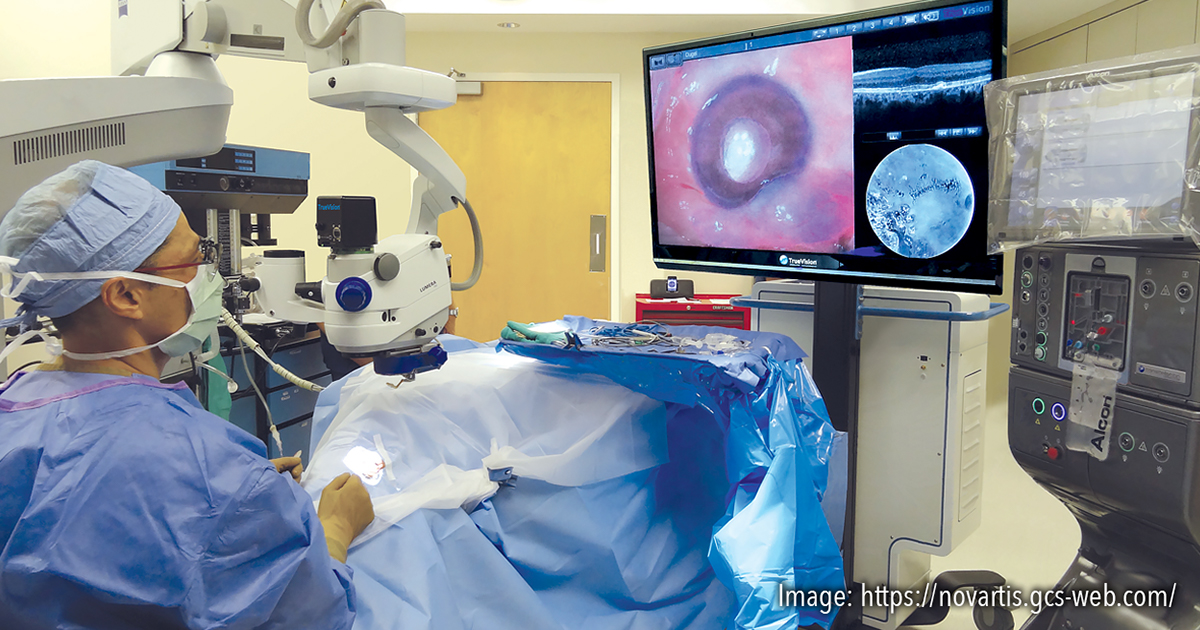
Ngenuity 3D consists of a 3D stereoscopic, high-definition digital video camera and workstation for providing magnified stereoscopic images of objects during microsurgery. TrueVision believes it will change the operating paradigm from using a microscope to performing heads-up surgery.
The system allows surgeons to operate looking up at a high-definition 3D monitor, instead of bending their necks to look through the eye-piece of a microscope, providing retinal surgeons unprecedented 3D visualization of the back of the eye with greater depth and detail during surgery than traditional microscopes.
Among the key advantages of the system that Dr. Tripathi notes are:
- a greater depth of field, allowing for a larger working space at the retina surface, in high magnifications;
- high-dynamic range imaging that allows the surgeon to reduce glare of instruments, and also to see into the shadows;
- use of digital image filtering, which can give different color effects;
- reduction of light levels by 50% or more, reducing light toxicity to both patient and surgeon; and
- better ergonomics, potentially reducing back and neck problems.
Real-World Experience
At the 2018 ASRS meeting, retina specialist Bozho Todorich, MD, PhD, described using the Ngenuity system for a novel technique of scleral indentation and transillumination for single-surgeon, unassisted vitrectomy and vitreous base shaving. The presentation was based on a recently published study in which the technique was utilized in six eyes of six patients during vitrectomy surgery for common vitreoretinal surgical diagnoses.1
In the study, the visualization of the vitreous cavity was digitally enhanced using the Ngenuity system, with the light amplification settings increased to near-maximal gain. Two independent surgeons viewed the case along with the primary surgeon and evaluated the surgical view. They found the view with scleral transillumination to be adequate in five of the six cases. In the remaining case, vitrectomy was completed using traditional endo-illumination and scleral depression. There were no intraoperative complications, and lighter fundus pigmentation, myopia, thin sclera, and absence of dense peripheral media opacities were associated with improved view with scleral transillumination.
For questions about this article, please contact Steve Lenier at Steve@stevelenier.com.
Reference
1. Todorich B, Stem MS, Hassan TS, Williams GA, Faia LJ. Scleral transillumination with digital heads-up display: a novel technique for visualization during vitrectomy surgery. Ophthalmic Surg Lasers Imaging Retina. 2018;49:436–439.